Congratulations, you are half way along in your pregnancy!
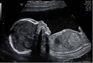
A routine ultrasound examination is offered to most patients at 20 weeks, the purpose of this examination is to assess:
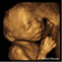
- Your baby’s anatomy. This involves a detailed examination of your baby’s brain, face, spine, heart, abdomen, renal tract, arms, legs, hands, feet and gender.
- Position of the placenta.
- Amniotic fluid volume.
- Pelvic anatomy and your cervix.
From 20 weeks, we do what we call a Morphology scan, this examination is expected to detect the main major fetal malformations, if any. However, it is important to understand, that we cannot detect all abnormalities. Some congenital heart abnormalities are progressive and unable to be detected at the 20 week ultrasound, cerebral palsy, biochemical abnormalities and some chromosomal abnormalities also cannot be detected.
The view of your baby may be limited by the:
Baby’s position at the time of examination, every attempt is made to move your baby for a better picture but occasionally you may have to return at a later date to complete the examination.
Tissue interposed between the ultrasound probe and the baby (fatty tissue or fibroids) sometimes this fatty tissue can absorb the ultrasound waves. If we are unable to obtain the right diagnostic images you may need to come back at a later date to complete the assessment.
Please inform the sonographer at the beginning of the ultrasound if you do not want to know the gender of your baby.